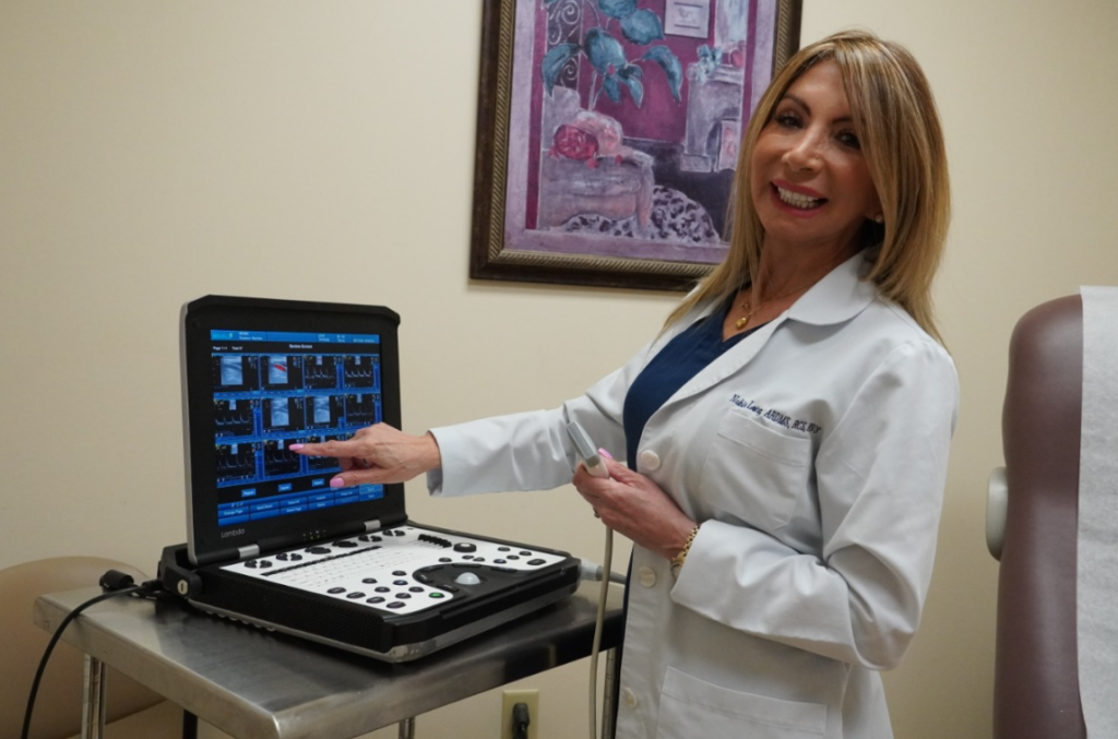
Nubia Long
tampa cardiovascular sonographer
Hi, I’m Nubia Long. I have more than eighteen years of experience in cardiovascular and abdominal testing.
Cardiovascular Mobile Ultrasound Inc. (CMU) is the provider of choice for diagnostic ultrasound services in the Tampa Bay area. CMU provides the convenience of serving the community in our offices and also by traveling to clinics, assisted living facilities (ALFs), and nursing homes to provide needed diagnostic ultrasound services. Our professional registry sonographers are reliable and caring. Reports will be available within 24-48 hours or immediately if requested. If you need diagnostic ultrasound services in your area, please don’t hesitate to contact us at Cardiovascular Mobile Ultrasound Inc. right away.
Medical credentials:
- B.S., ARDMS., RCS., RVS.
- Diagnostic Ultrasound School
- Registered Diagnostic Medical Sonographer - (American Registry for Diagnostic Medical Sonography)
- Registered cardiac sonographer. Cardiovascular (International Accreditation - CCI)
- Registered Vascular Technologist- CCI
Our
Services
HIFU
If you’re looking for a non-invasive way to rejuvenate your skin, you won’t want to miss this. HIFU, or High Intensity Focused Ultrasound, is a cutting-edge treatment that uses ultrasound technology to lift and tighten the skin, giving you a more youthful and radiant appearance.
Unlike traditional cosmetic procedures that require surgery, injections, or other invasive methods, HIFU is completely non-invasive and requires no downtime. You can have the treatment done during your lunch break and be back to your normal activities in no time.
So, what makes HIFU so effective? It works by delivering focused ultrasound energy to the deeper layers of the skin, where it heats the underlying tissue and stimulates the production of new collagen. This results in a firmer, more elastic skin that looks and feels younger.
But don’t just take our word for it! HIFU has already become a popular choice for many people looking to rejuvenate their skin and achieve a more youthful appearance. And with no scars, bruises, or long-term effects, it’s a safe and effective way to achieve your beauty goals.
Contact us today to learn more and schedule your appointment.
Text or Call 813-417-5888.
Before
After
Before
After
Before
After
Previous
Next
SCREENING EXAMS
Preventive & Affordable healthcare near you
Cmu is conducting better Cardiovascular healthcare making it convenient and affordable to you, by offering regular preventive screenings. With the advanced ultrasound technology radiologist and cardiologist are able to see inside your heart, arteries, and organs to obtain more information about your health.
Cmu is now bringing this valuable service to your neighborhood.
In less than one hour, you can get a complete cardiovascular screening in the comfort and privacy of our clinic. With just one appointment, technologists can perform a series of test which look at the major risk factors for :
- Family history of heart attack
- Stroke
- Aneurysm
- Vascular desease
- High blood pressure
- Diabetes
- High Cholesterol
Show More
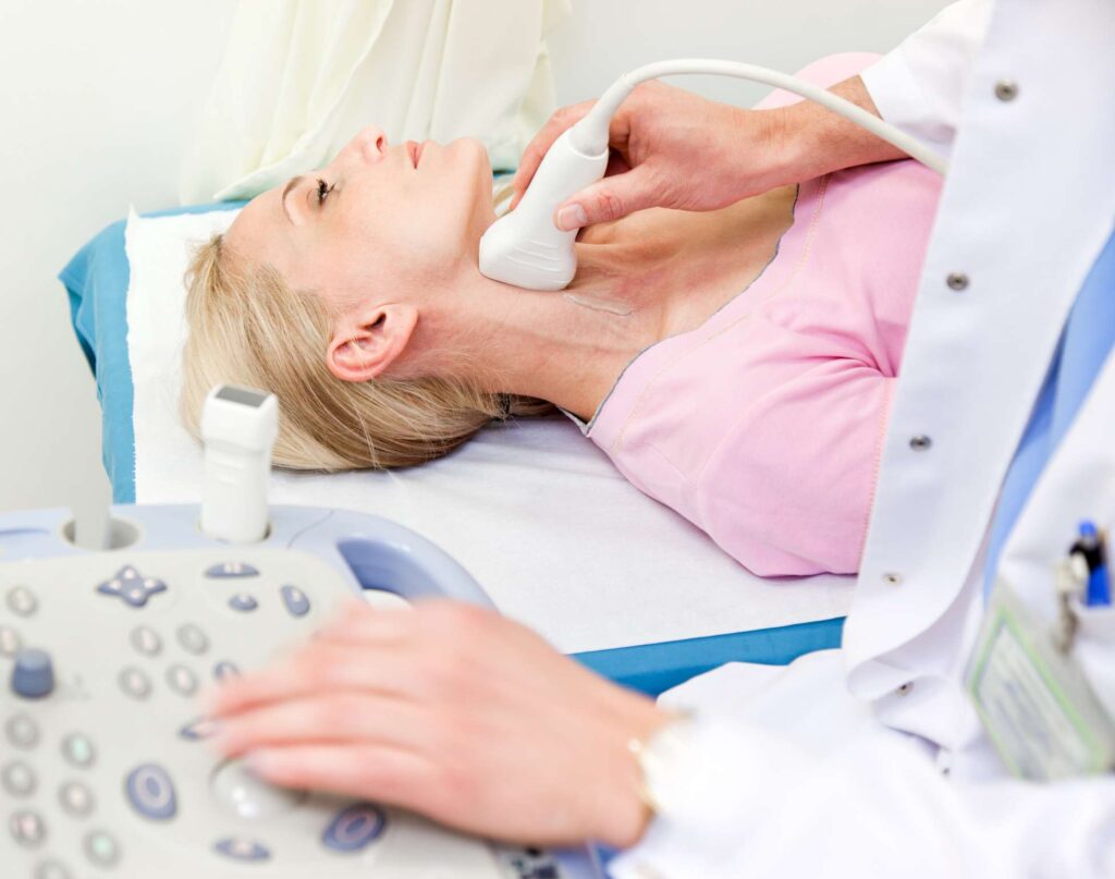
Carotid ultrasound
Cardiovascular Mobile Ultrasound Inc. (CMU) is the provider of choice for diagnostic ultrasound services in the Tampa Bay area. CMU provides the convenience to serve the community at our offices and also by traveling to clinics, assisted living facilities (ALFs), and nursing homes to render diagnostics ultrasound services needed. Our professional register sonographers are reliable and caring. Reports will be available within 24-48 hours or stat if requested. If you need diagnostics ultrasound services in your area, don’t hesitate to contact us at Cardiovascular Mobile Ultrasound Inc. right away.
Show More
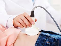
Abdominal Aorta Ultrasound Imaging
Ultrasound of the abdominal aorta is primarily used to evaluate for an aneurysm which is an abnormal enlargement of the aorta usually from atherosclerotic disease.
When the walls of the abdominal aorta become weak, they may balloon outward. If the aorta reaches over 3 centimeters in diameter, it is then called an abdominal aortic aneurysm (AAA). As the aneurysm gets larger, the risk of rupture increases.
Ultrasound imaging of the aorta is useful for measuring its size to screen for AAA. Screening is particularly recommended for men over the age of 60 who have ever smoked and for anyone with a family history of AAA. In addition to screening, ultrasound is also a useful tool after the diagnosis of AAA to monitor its size on a regular basis to see if it needs to be repaired.
Show More
Thyroid ultrasound
Ultrasound of the thyroid produces pictures of the thyroid gland and the adjacent structures in the neck. The thyroid gland is located in front of the neck just above the collar bones and it’s shaped like a butterfly, with one lobe on either side of the neck connected by a narrow band of tissue. It is one of nine endocrine glands located throughout the body that make and send hormones into the bloodstream.
Why should you have a thyroid ultrasound.
- it determines if a lump in the neck is arising from the thyroid or an adjacent structure
- analyzes the appearance of thyroid nodules and determines if they are the more common benign nodule or if the nodule has features that require a biopsy.
- looks for additional nodules in patients with one or more nodules felt on physical exam
- to see if a previously found thyroid nodule has substantially grown over time
Show More
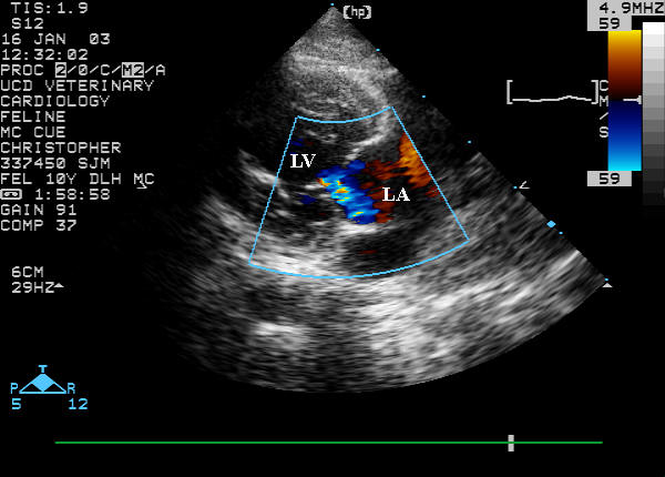
Echocardiogram imaging
An echo uses sound waves to create pictures of your heart’s chambers, valves, walls and the blood vessels (aorta, arteries, veins) attached to your hear
How to prepare for an Echocardiogram.
No special preparations are necessary for a standard transthoracic echocardiogram. You can eat, drink and take medications as you normally would.
What can be seen from an Echo ultrasound.
- The size and shape of your heart, and the size, thickness and movement of your heart’s walls.
- How your heart moves.
- The heart’s pumping strength.
- If the heart valves are working correctly.
- If blood is leaking backwards through your heart valves (regurgitation).
- If the heart valves are too narrow (stenosis).
- If there is a tumor or infectious growth around your heart valves.
The test also will help the doctor find out if there are
- Problems with the outer lining of your heart (the pericardium).
- Problems with the large blood vessels that enter and leave the heart.
- Blood clots in the chambers of your heart.
- Abnormal holes between the chambers of the heart.
Show More
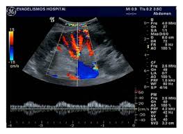
Renal Doppler ultrasound
The Renal Artery Doppler Ultrasound uses high frequency sound waves to assess the blood flow into and out of the kidneys.
If you have uncontrolled high blood pressure, your doctor may request this exam to determine if there is a narrowing in the blood vessels leading to each kidney from the aorta. Tiny blood vessels within each kidney are also evaluated.
Show More
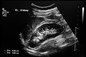
Renal (kidney) ultrasound
Renal ultrasonography is the examination of one or both kidneys using medical ultrasound. Ultrasonography of the kidneys is essential in the diagnosis and management of kidney-related diseases. The kidneys are easily examined, and most pathological changes in the kidneys are distinguishable with ultrasound
A kidney ultrasound may be used to assess the size, location, and shape of the kidneys and related structures, such as the ureters and bladder. Ultrasound can detect cysts, tumors, abscesses, obstructions, fluid collection, and infection within or around the kidneys.
Indications for a Renal Ultrasound
- Flank Pain
- Hematuria
- Kidney Stones
- Proteinuria
- Hydronephrosis
- Bladder Infection
- Kidney Infection
What Ultrasound Can Determine
- Kidney Stones
- Hydronephrosis
- Masses or Cyst
- Dilated Ureter(s)
- Non-excavation of the Bladder
- Renal Carcinoma
- Acute Renal Failure
Show More
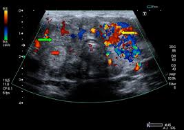
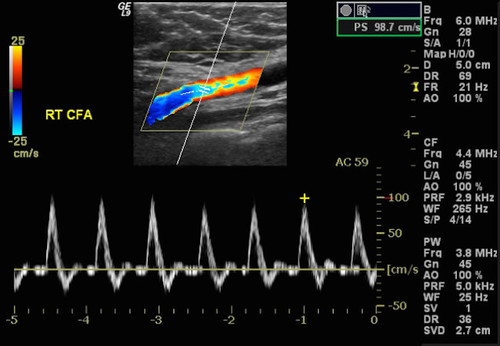
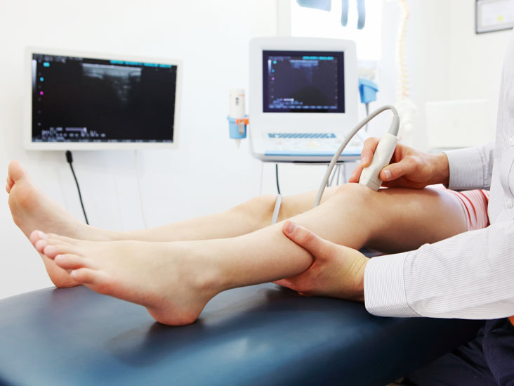
Arterial Duplex/Doppler
Ultrasound is a procedure that uses sound waves to “see” inside your body. An arterial duplex ultrasound uses sound waves to create a color map of the arteries in your legs to identify:
- Narrowing of your vessels that may be causing leg pain when walking
- Resting leg pain
- Foot, ankle, heel or toe ulcers
- Skin discoloration
Ultrasound duplex scanning can provide additional information that may guide therapeutic decisions. The location and severity of arterial narrowings and occlusions can be identified. The vascular technologist can map disease in lower-extremity segments with great accuracy.
The additional information from duplex scanning can help determine if arterial disease might be appropriately treated with an endovascular intervention. The type of treatment and the technical approach can be guided by the duplex scan findings. Information from the duplex scan can help patients be better informed about their options. Duplex scanning after intervention can provide objective information about the success of the procedure and serial follow-up examinations can identify recurrent problems at an early stage, sometimes prompting follow-up interventions.
A Doppler ultrasound uses sound waves to produce images that highlight blood flow in the leg arteries. This test detects and evaluates any blockages caused by plaque buildup.
An average of 1 in 20 people over the age of 50, or as many as 10 million Americans, suffer from peripheral arterial disease. This disease is caused by atherosclerosis, the hardening of arteries. The slow process of plaque buildup on the artery walls over time causes a narrowing of the vessel walls, decreasing the blood flow to the muscles in the legs. A simple Arterial Duplex Ultrasound will detect this plaque in its early stages and prevent further damage.
Peripheral vascular disease, if undetected, may lead to gangrene and ultimately amputation or death. The Upper or Lower Extremity Arterial Ultrasound is a noninvasive, simple 30-minute exam that can prevent this.
Indications for an Arterial Ultrasound
- Leg Pain
- Rest Pain
- Abdominal Aneurysm
- Pre/Post Surgery
- Diabetes
- Patient is a Smoker
- Non-Healing Ulcer
- Claudication
What Ultrasound Can Determine
- Peripheral Vascular Disease (PVD)
- Popliteal Aneurysm
- Bakers Cyst
- Stenosis
- Arterial Blockage
Show More
Venous Doppler Ultrasound
A venous Doppler ultrasound is a diagnostic test used to check the circulation in the large veins in the legs (or sometimes the arms). This exam shows any blockage in the veins by a blood clot or “thrombus” formation.
This test involves no needles, catheters or dye. Ultrasound is used to listen to the flow of the blood through your veins.
Normal Circulation
The veins return blood to the heart. There are two sets of veins in the legs:
- Deep veins – which are underneath the leg muscles
- Superficial veins – which are right underneath the surface of the skin
The veins in the leg deflate easily; blood flows under low pressure and against gravity. Two things help the blood pump more efficiently:
- The calf “muscle pump.” As you walk, calf muscle movement pushes blood upward.
- Venous valves – the one-way valves in the veins that prevent blood from flowing back into your feet.
- DVT (deep venous thrombosis)is a serious health concern, because it is possible that the clot could break off and travel to your lungs. It is then referred to as a pulmonary embolus. The risk of pulmonary embolus is decreased if you recognize the symptoms of a DVT quickly and get it treated right away.
- Superficial thrombophlebitis is a clot that has formed in the veins near the surface of your skin. Clotting can occur after injury or in old varicose veins. Superficial thrombophlebitis usually responds very well to local treatment.
- Chronic venous insufficiency (CVI)
- is a condition that occurs when the venous wall and/or valves in the leg veins are not working effectively, making it difficult for blood to return to the heart from the legs. CVI causes blood to “pool” or collect in these veins, and this pooling is called stasis.
Thrombophlebitis
Is an inflammatory process that causes a blood clot to form and block one or more veins, usually in your legs. The affected vein might be near the surface of your skin (superficial thrombophlebitis) or deep within a muscle (deep vein thrombosis, or DVT).
Risk Factors
No one understands completely why some people are more likely than others to develop a DVT. Some situations increase the chance of clots forming in the deep veins:
- Surgery or injury
- Long-term bedrest or immobility
- Smoking
- Obesity
- A history of venous thrombosis
- Pregnancy
Diagnosis
If your doctor thinks you might have a venous disorder, he or she may order a diagnostic exam to determine what type of a problem you have and how severe it might be.
Show More
Insurance
Payment methods


Contact
- 813-962-8350
- 813-417-5888
- 866-215-7823
- nubialong@cmultrasound.com
Copyright © cmultrasound 2021